- June 2014
- Volume 15
- Issue 6
Early Results Suggest Tomosynthesis Marks Major Advance in Breast Imaging
The first time Christina T. Giuliano, MD, saw a digital mammogram, she knew the extra clarity and detail would profoundly improve the diagnostic process.
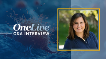
Christina T. Giuliano, MD
Director, Breast Imaging
Maimonides Medical Center
Brooklyn, NY
The first time Christina T. Giuliano, MD, saw a digital mammogram, she knew the extra clarity and detail would profoundly improve the diagnostic process.
She had the same feeling when she first examined images produced by digital tomosynthesis, and early research has supported her instinct that another profound improvement is on the way.
“Studies to date have all found large increases in cancer detection rate and large decreases in the rate of false-positive recalls,” said Giuliano, director of Breast Imaging at Maimonides Medical Center in Brooklyn, during an interview.
“If ongoing trials confirm those findings, tomosynthesis will establish itself as the biggest advance in breast imaging since digital replaced film, and it will rapidly become a new standard of care,” she said. “Anyone who treats breast cancer should understand some basic things about it.”
Giuliano explained those basics, starting with the technology, in a presentation during the 31st Annual Miami Breast Cancer Conference® that Physicians’ Education Resource, LLC (PER®) hosted in Florida in March.
Digital tomosynthesis differs from standard mammography in much the same way as computed tomography (CT) scans differ from X-rays.
Rather than take individual pictures with a stationary X-ray tube, it takes a series of exposures from a tube that arcs around the patient. Physicians then examine those images “slice by slice.”
Tomosynthesis involves far fewer exposures than a CT scan and far less radiation. Tomosynthesis plus a standard mammogram involves about twice as much radiation as a standard mammogram alone. Synthesized 2D images may soon replace the standard 2D views and, if that happens, the radiation figure should drop to values similar to a standard mammogram alone.
“To put the radiation risk in perspective for patients, I tell them that the additional exposure is about equivalent to the exposure they get from the sun in a month,” said Giuliano. “It really is a very small amount of radiation.”
Giuliano contrasted those small risks with the far larger benefits reported across the published literature.
Early results from the Oslo Tomosynthesis Screening Trial, for example, found that a combination of tomosynthesis and standard mammography increased tumor detection by 40% and decreased the false detection rate by 15%. Other results from Europe, where regulators approved tomosynthesis in 2008, three years ahead of the FDA, have been nearly as promising.
That said, Giuliano warns that differences in patient populations and standards of care may prevent exact experiences abroad from translating directly to the United States.
“Sticking with the Oslo example, Americans who read the study need to remember that Norwegians only get mammograms every other year, which could certainly affect the magnitude of the cancer detection results,” Giuliano said.
“Also, the Europeans traditionally have a lower rate of false-positive recalls than we do, so American practices might see even more benefit from tomosynthesis on that front.” Studies under way in the United States will eventually provide domestic figures on tumor detection and false positives, but it may take several more years for even the oldest European studies to determine whether or not tomosynthesis extends lifespan.
Still, even without lifespan data, many American physicians and patients have decided that increased tumor detection and decreased false positives justify not only the extra radiation but also a number of extra costs. The machine itself runs about $100,000 more than an equivalent model that only performs 2D digital mammograms. Moreover, all those extra images that make tomosynthesis such a powerful diagnostic tool increase costs at other levels.
“The more images you have, the more time it takes for the radiologist to examine everything,” Giuliano said.
Studies suggest that it initially takes radiologists several times longer to review each patient’s images when they begin working with tomosynthesis but that, with experience, their speed increases and they require about twice as much time per patient.
Giuliano’s personal experience, which includes a first-hand taste of what it’s like to work with tomosynthesis, suggests that it takes quite some time to reach maximum speed. Giuliano has been using tomosynthesis for about two years now, and it still takes her nearly three times as long to interpret all the images.
In addition to requiring more time for interpretation, all those extra images require more space on the servers that will store them and more computing power from the office’s system.
Solutions for data transfer and storage, Giuliano warns, will involve your IT department.
“These are big files, and there are a lot of them. They are going to burden your computers, and they’re going to cost real money to store,” Giuliano said. “My medical center is spending an extra $20,000 to $30,000 per year for image storage above what I’d need for 2D mammograms.”
Worse, those costs are not offset by higher reimbursements from insurance companies or the government. Third-party payers have yet to write codes for tomosynthesis, let alone start paying extra for either the equipment or the extra time and effort that the technology requires of doctors.
Among those practices that offer it, some bill customers $50 or $100 to compensate for the added costs, while many others offer it free to patients who also get standard mammograms.
“The financials sound scary, but Yale, which is charging nothing for tomosynthesis, did a study that found the service may save money by lowering diagnostic recalls,” Giuliano said.
Articles in this issue
over 11 years ago
CLL Researcher Excited About Outlook for CDK Inhibitorsover 11 years ago
Novel Therapies, Strategies in Development for NSCLC


