Trilaciclib in SCLC: Mechanism of Action and Dosage
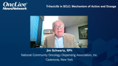
Jim Schwartz, RPh, provides key insights into the recent approval of trilaciclib and discusses how CDK4/6 inhibition potentially improves response to cytotoxic chemotherapy by protecting patients from myelosuppression.
Jim Schwartz, RPh: I want to start with the role of CDK4/6 in normal bone marrow function. CDK4/6 is cyclin-derived kinase. There are a number of them in the body, 1 through 9. These are specifically CDK4 and CDK6, and they form complexes in the hematopoietic cells. They drive cell proliferation. It’s important for the cell cycle for these 2 kinases to be present. In the G1 phase, they bind to the D-cyclin protein. As that builds up, as more of those conjugates form, it triggers a positive feedback loop that commits cells so they pass from the restriction point, from the G1 portion of the cell cycle to the S transition, where DNA synthesis starts. Both kinases are already active and present in the hematopoietic cells. At that point in the G1 cycle, they begin combining with the D-cyclin. They build up. That causes at a certain point for it to move on to the S phase. That’s the reason why these 2 are important substances in the maturation of these cells.
Now that we know the role of the CDK4 and CDK6 kinases, we want to talk about a new agent that’s out now to inhibit their action, and that’s trilaciclib. It’s a small-cell molecular inhibitor of these 2 kinases, and it protects the hematopoietic cells from chemotherapy-induced exhaustion. When they’re introduced into the body, they enter the hematopoietic cells, and they bind to the CD4 and CDK kinases. Because they’re tying up those 2 kinases, they cannot bind to the D-cyclin. All those are tied up, so the cell does not reach the point where they have enough CDK4/6 and D-cyclin conjugates to enter the S phase. They are stuck in the G1 phase, and they cannot move on to S or the DNA synthesis or the G2, where they have further growth, and certainly not to mitosis. As long as these drugs are inhibited by this new drug trilaciclib, the cell is stuck in the G1 portion of the cell cycle, and that protects it from anything that might occur. Chemotherapy acts in many places: the S, the G2, and the M phase. Because it never enters those, they’re protected from being damaged or even destroyed by the chemotherapy. This cell cycle rest by trilaciclib is transient, reversible, and dose related. As you know, there are a number of oral CDK4/6s available. Why are they not working in this way? The primary answer is that it’s dose related. Because they do this, it protects the cell and provides lasting protection against short- and long-term toxicity that could be caused by these hematopoietic cells.
We have the mechanism of action of trilaciclib. How does this benefit a patient undergoing chemotherapy for small-cell lung cancer? The regimens being given—etoposide and carboplatin with or without an immunotherapy, as well as topotecan in the second or third line—are very myelosuppressive. By preventing the cells from being injured during the administration of the chemotherapy, it allows the patient to stay on the prescribed dose of the chemotherapy longer. When they develop neutropenia or anemia or any of these things, there have to be dose stoppages. They may have to reduce the dose. It avoids treatment delays and the expensive treatment of myelosuppressive adverse effects that occur, such as neutropenia, infection, and thrombocytopenia. These things can lead to the risk of infection, fatigue, and bleeding. Overall, it allows the patient to stay on the recommended dose longer, avoid delays, and avoid the treatment of adverse effects. Ultimately, the purpose of cytotoxic chemotherapy is to get as much drug to the patient as you can without too many adverse effects. The addition of trilaciclib cuts out most of the hematopoietic damage and allows the patient to get more of the drug and stay on it longer, resulting in better outcomes.
How is this drug given? The dosing is 240 mg/m2. It’s given within 4 hours of chemotherapy. It’s an irritating drug. But it has effect within 4 hours, so it has to be given within 4 hours of the chemotherapy administration. It reaches its maximum effect at 12 hours and has a half-life of 36 hours, so it acts a long time but goes away quickly. It should be administered in normal saline. It needs to be at a concentration of 0.3 to 3 mg/mL. The drug is irritating, so it needs to be diluted. In general, 250 mL should take care of most doses since it’s in 300-mg vials and the dose is 240 mg/m2. Most patients are going to need more than 300 mg/m2. It will probably be between 300 and 400 or 500 mg/m2. If you put it in 250 mg/m2, that will get it within the range of 0.3 to 3 mg/mL. It should have an inline filter, 0.2- or 0.22-micron filter inline. It can be given over 30 minutes, and it would be good for the first dose to start out slowly. It’s an irritant, and it can cause injection site reactions. This is seen more with a peripheral IV [intravenous] administration. It should not be as much of a problem given through a central line. But when it’s given through a peripheral IV, then it needs to be watched carefully, and the arm that it’s given in should be rotated each time so 1 vein is not worn out from getting this drug.
Transcript Edited for Clarity


